
 Submit Manuscript
Research Article | Open Access
Predictors of Tumor Extent and Clinically Significant Incidental Prostate Cancer in Men with PSA <4 ng/mL Undergoing Transurethral Resection of the Prostate in North-West Cameroon
Tagang Titus Ngwa-Ebogo1,2,3, Forcha Yannick Tandu4, Marie Louise Manka’a2, Azamafac Kareen Esenwoh1,3, Marcella Derboise Christelle Biyouma5, Landry Oriol Mbouche6, Fru Forbushi Angwafo III6
1Faculty of Health Sciences, The University of Bamenda, Bamenda, Cameroon.
2MD Research Group, Bamenda, Cameroon.
3Regional Hospital Bamenda, Bamenda, Cameroon.
4University of Abuja Teaching Hospital, Abuja, Nigeria.
5Urology Service, Hôpital Laquintinie, Douala, Cameroon.
6Faculty of Medicine and Biomedical Sciences, University of Yaoundé I, Yaoundé, Cameroon.
Correspondence: Ngwa-Ebogo Titus Tagang (Faculty of Health Sciences, The University of Bamenda, Bamenda, Cameroona; Email: ngwa.ebogo@uniba.cm).
Annals of Urologic Oncology 2026, 9: 7. https://doi.org/10.32948/auo.2026.10.11
Received: 04 Apr 2026 | Accepted: 21 Oct 2026 | Published online: 11 Nov 2026
Methods This retrospective two-center case series included 103 men with PSA <4 ng/mL who underwent TURP for presumed benign prostatic hyperplasia between January 2020 and July 2024 and had unsuspected adenocarcinoma. We reviewed clinical and histopathological records. Exploratory multivariable logistic regression assessed factors associated with T1b disease and International Society of Urological Pathology Grade Group ≥2 cancer. The case-only design precluded estimation of iPCa frequency among all TURP procedures.
Results Mean age was 67.0 ± 6.9 years, mean PSA was 2.23 ± 0.59 ng/mL, and mean PSA density (PSAD) was 0.037 ± 0.016 ng/mL/mL. T1b disease occurred in 41 of 103 patients (39.8%), and 43 (41.7%) had Grade Group ≥2 cancer. In adjusted analyses, PSAD showed a borderline association with T1b disease (OR 2.66 per 0.01 ng/mL/mL, p = 0.050), while absolute PSA was inversely associated with T1b disease (OR 0.17 per ng/mL, p = 0.041). Higher preoperative International Prostate Symptom Score was associated with Grade Group ≥2 cancer (OR 1.08 per point, p = 0.024). Model discrimination was modest (AUC 0.66 for T1b and 0.65 for Grade Group ≥2).
Conclusion Substantial proportions of men with iPCa and PSA <4 ng/mL had T1b or Grade Group ≥2 disease. These findings support histopathological evaluation of TURP tissue and structured assessment after incidental diagnosis. Associations with PSAD and symptom burden require external validation before clinical use.
Key words Incidental prostate cancer, Transurethral resection of the prostate, PSA density, ISUP grade group, Cameroon
Preoperative risk assessment remains difficult because PSA is prostate-specific but not cancer-specific. PSA may be increased by benign prostatic enlargement, prostatitis or other inflammation, urinary retention, recent catheterization, and instrumentation; conversely, a low PSA does not exclude aggressive or relatively low-PSA-producing tumors. Before TURP, discordance between PSA and gland size is therefore clinically important: a relatively high PSA in a small prostate, or a PSA value disproportionate to the gland volume, should prompt closer evaluation for occult cancer when digital rectal examination, MRI, and biopsy are available. PSA density (PSAD), calculated by dividing total PSA by prostate volume, expresses PSA production relative to gland size and often discriminates clinically significant disease better than absolute PSA alone [5, 6]. A PSAD threshold near 0.15 ng/mL/mL is frequently used to support biopsy or MRI decisions, although the optimal threshold varies with prostate volume, population, and diagnostic pathway [7, 8]. Composite laboratory indices have also been explored as adjunctive predictors, but their evidence base remains preliminary [9]. The hemoglobin-albumin-lymphocyte-platelet (HALP) score has shown preliminary discriminatory value for incidental and higher-grade prostate cancer in a single-center TURP study; however, external validation is required before it can be incorporated into routine risk stratification [9].
The likelihood of detecting iPCa also depends on the extent of tissue removal and histopathological examination. More complete embedding of TURP chips increases the probability of identifying small malignant foci [10]. Nevertheless, the absolute weight of resected tissue is an imperfect surrogate for cancer burden because T1a/T1b staging is based on the proportion of the resected specimen involved rather than on the proportion of the entire prostate containing tumor. A small cancer focus may represent ≥5% of a limited resection and therefore be classified as T1b, whereas the same absolute tumor volume may be diluted within a larger resection. For this reason, the ratio of resected tissue weight to estimated total gland weight may provide more meaningful context for estimating the “incidental” cancer burden and reduce distortion caused by very small or very large resections [11, 12]. The relationship among gland size, resection fraction, tumor distribution, and pathological sampling is therefore complex and should not be reduced to specimen weight alone.
Contemporary data from sub-Saharan Africa remain limited. PSA screening is inconsistent, access to MRI and image-guided biopsy is restricted, and many men present with severe lower urinary tract symptoms requiring surgical treatment [4, 13, 14]. In this context, TURP may function both as treatment for obstruction and as an important diagnostic opportunity. Existing African reports have primarily estimated the frequency of carcinoma in TURP specimens or compared cancer-positive and benign specimens, while fewer studies have examined the factors associated with extent and grade after incidental cancer has already been identified. The distinction is clinically important: predictors of whether cancer will be found are not necessarily the same as predictors of whether an identified cancer is T1b or Grade Group ≥2. To our knowledge, no previous two-center study from Cameroon has focused specifically on men with PSA <4 ng/mL who were subsequently found to have previously unsuspected adenocarcinoma at TURP. The present study is novel in separately evaluating factors associated with pathological tumor extent (T1b) and clinically significant grade (Grade Group ≥2) within this low-PSA population. It also evaluates PSAD and a standardized symptom score using variables that are obtainable in hospitals where advanced imaging may be unavailable. We aimed to characterize the clinicopathological profile of these cancers and hypothesized that PSAD, symptom severity, and other preoperative variables might provide information beyond absolute PSA alone. Because only cancer-positive cases were available, the study was designed to describe tumor characteristics and predictors rather than to calculate the local incidence of iPCa among all TURP procedures.
We conducted a retrospective two-center case series using medical records from Bamenda Regional Hospital and Nkwen Baptist Hospital in the North-West Region of Cameroon. Eligible patients were men who underwent monopolar or bipolar TURP between January 2020 and July 2024 for symptomatic presumed benign prostatic hyperplasia, had a preoperative total PSA <4 ng/mL, and were subsequently found to have prostatic adenocarcinoma in the TURP specimen. Men with a known diagnosis of prostate cancer before TURP, a previous prostate biopsy or prostate operation, or current 5-alpha-reductase inhibitor therapy were excluded to focus on true incidental cancers. Only records containing sufficient clinical and pathological information for staging and grading were analyzed. The analytic dataset did not include a reliable denominator of all eligible TURP procedures; consequently, it could not be used to estimate the incidence of iPCa. Ethical approval and administrative authorizations were obtained, and the institutional review board waived individual informed consent for this retrospective analysis of de-identified records.
Data collection
We collected demographic characteristics including age, marital status, educational level, occupation, and body mass index, together with medical history such as hypertension, diabetes mellitus, tobacco and alcohol exposure, and family history of prostate cancer. Lower urinary tract symptom severity was assessed using the physician-administered International Prostate Symptom Score (IPSS). Digital rectal examination findings were categorized as benign-feeling/normal or enlarged/asymmetric without a discrete malignant nodule. Total serum PSA was measured before TURP using routine immunoassay techniques. Prostate volume was estimated by transrectal ultrasonography with the ellipsoid formula (length × width × height × 0.52), and PSAD was calculated as total PSA divided by prostate volume in ng/mL/mL. Although a PSAD threshold around 0.15 ng/mL/mL is commonly used in biopsy and MRI pathways, PSAD was primarily analyzed as a continuous variable because values in this deliberately low-PSA cohort were generally far below that threshold. Multiparametric MRI was not obtained preoperatively because of limited availability and affordability; therefore, PI-RADS scores or other radiological features could not be correlated with pathological stage or grade.
Histopathological examination of the TURP specimens was performed by hospital pathologists according to routine institutional protocols. TURP chips were fixed in formalin and all available tissue was submitted for embedding and microscopic assessment whenever feasible. Pathology reports were reviewed for adenocarcinoma, Gleason score, ISUP Grade Group, and the estimated proportion of resected tissue involved by carcinoma. Tumors involving <5% of the resected tissue were classified as T1a, while those involving ≥5% were classified as T1b [15]. Clinically significant prostate cancer was defined a priori as ISUP Grade Group ≥2 (Gleason score ≥3+4). The principal outcomes were T1b disease and Grade Group ≥2 cancer. Resected tissue weight, the ratio of resected tissue weight to total estimated gland weight, high-resolution archival photomicrographs, post-TURP MRI or confirmatory biopsy findings, radical prostatectomy pathology, and longitudinal treatment or survival outcomes were not consistently available and were therefore not analyzed. No unrecorded pathological, imaging, or follow-up information was inferred. Free-to-total PSA ratio and high-grade prostatic intraepithelial neoplasia (HGPIN) were not consistently documented in the source records and were therefore not analyzed.
Statistical analysis
Continuous variables were summarized as mean ± standard deviation or median with interquartile range, as appropriate, and categorical variables as frequencies and percentages. Baseline characteristics were compared between T1a and T1b groups using Student’s t test or the Mann–Whitney U test for continuous variables and the chi-square test or Fisher’s exact test for categorical variables. Exploratory multivariable logistic regression models evaluated associations with (1) T1b disease and (2) Grade Group ≥2 cancer. Candidate clinical variables included age, total PSA, prostate volume, PSAD, and preoperative IPSS. Percentage tissue involvement was not interpreted as a predictor of T1b because it defines the T1a/T1b outcome; it was considered only in the Grade Group ≥2 model. All clinically selected variables were entered simultaneously rather than selected by automated stepwise procedures. PSAD odds ratios were rescaled to a clinically interpretable 0.01 ng/mL/mL increment because a one-unit change is far outside the observed range and would generate an unhelpfully large odds ratio. Because PSAD is mathematically derived from PSA and prostate volume, models containing all three variables were treated as exploratory and estimates were interpreted cautiously for possible multicollinearity, coefficient instability, and suppression effects. Logistic regression results are presented as odds ratios with p values, and 95% confidence intervals where available. Model discrimination was assessed using the area under the receiver operating characteristic curve (AUC), and explanatory power using pseudo R². These metrics were used to describe model performance rather than to claim a validated clinical prediction tool. A two-sided p<0.05 was considered statistically significant. Analyses were performed in Python using Statsmodels and cross-checked in R.
A total of 103 men met the inclusion criteria. Table 1 summarizes their clinicopathological characteristics overall and by incidental stage. The mean age was 67.0 ± 6.9 years (range 52–82), and 70% were aged 60 years or older. Most participants were married (69%), and approximately 39% had secondary education or higher. Hypertension was present in 56% and diabetes mellitus in 13%. Fifteen percent were current or former smokers, while 41% reported regular alcohol use. The mean preoperative IPSS was 17.1 ± 6.8, indicating predominantly moderate-to-severe lower urinary tract symptoms. These characteristics describe the cancer-positive case series and should not be interpreted as the distribution among all men undergoing TURP during the study period (Table 1).
On digital rectal examination, 73 men (70.9%) had a benign-feeling prostate with no palpable nodule, while 30 (29.1%) had diffuse enlargement or asymmetry without a discrete malignant nodule. The mean transrectal ultrasound prostate volume was 65.3 ± 18.2 mL. Despite the deliberately low absolute PSA values (mean 2.23 ± 0.59 ng/mL), the mean PSAD was 0.037 ± 0.016 ng/mL/mL. These values were substantially below the commonly used 0.15 ng/mL/mL threshold, and PSAD was therefore evaluated as a continuous measure rather than interpreted as validation of a specific cutoff. The low average PSAD also illustrates why a threshold derived from biopsy or MRI cohorts cannot be transferred uncritically to a selected population in which all PSA values were below 4 ng/mL.
All 103 patients had adenocarcinoma detected on TURP histopathology. Sixty patients (58.3%) had ISUP Grade Group 1 disease, 28 (27.2%) had Grade Group 2, and 15 (14.6%) had Grade Groups 3–4; no Grade Group 5 tumor was recorded. Among the 41 T1b cases, 20 were Grade Group 1, 15 were Grade Group 2, and 6 were Grade Groups 3–4. Among the 62 T1a cases, 40 were Grade Group 1, 13 were Grade Group 2, and 9 were Grade Groups 3–4. The Grade Group distribution by incidental stage is presented in Table 2 and Figure 1. Clinically significant cancer, defined as Grade Group ≥2, was present in 43 patients (41.7%). It occurred in 22/62 T1a cases (35.5%) and 21/41 T1b cases (51.2%). Table 3 reports a discrepant cross-tabulation that requires verification against the source data. Although Grade Group ≥2 disease was numerically more common in T1b cases, the unadjusted association did not reach statistical significance (OR 1.91, 95% CI 0.85–4.26; p=0.115). This pattern demonstrates that limited involvement of the TURP specimen cannot be assumed to represent biologically insignificant cancer and that grade and stage describe related but distinct pathological dimensions. High-grade tumors were relatively uncommon overall, but the presence of Grade Groups 3–4 in both stage categories indicates that potentially important disease may be encountered even when the involved tissue constitutes <5% of the resection. Thus, both limited and more extensive TURP involvement contained clinically significant tumors, and T1a/T1b stage alone did not fully represent biological aggressiveness.
Incidental stage distribution
Overall, 62 patients (60.2%) were classified as T1a because carcinoma involved <5% of the resected tissue, and 41 (39.8%) were classified as T1b because involvement was ≥5%. Mean tumor involvement was approximately 2.3% in T1a cases and 19% in T1b cases. These percentages describe the resected TURP material and should not be interpreted as direct measurements of whole-gland tumor volume. The fraction of the gland removed during TURP was not consistently recorded, so the ratio of resected tissue weight to total gland weight could not be calculated. This is relevant because an identical absolute tumor focus may represent a larger percentage of a small resection than of a large resection.
Patients with T1a and T1b disease were broadly similar in baseline characteristics. Mean age was 67.0 versus 67.1 years, mean PSA was 2.30 versus 2.22 ng/mL, and mean prostate volume was 66.4 versus 63.7 mL in the T1a and T1b groups, respectively. Mean PSAD was also similar on unadjusted comparison (0.037 vs 0.038 ng/mL/mL), while mean IPSS was modestly higher in T1b cases (18.3 vs 16.4) without reaching statistical significance. These small differences show that the stage groups overlapped considerably in their preoperative clinical profiles. The higher proportion of Grade Group 2 tumors in T1b cases suggested a possible relationship between tissue extent and grade, but the overall Grade Group ≥2 comparison did not achieve statistical significance. No single preoperative clinical variable clearly separated T1a from T1b disease on simple comparisons, which provided the rationale for evaluating the variables jointly in exploratory regression models. Figure 2 retains the original exploratory visualization of PSAD and recorded tissue involvement; because percentage involvement defines incidental stage, the figure is descriptive and was not used to treat tissue involvement as an independent predictor of T1b disease. The plot should not be interpreted as a substitute for the pathology-derived stage criteria or as validation of a particular PSAD threshold.
|
Table 1. Baseline clinicopathologic characteristics of patients. |
|||
|
Parameter |
Overall (N=103) |
T1a (n=62) |
T1b (n=41) |
|
Age (years) |
67 ± 6.9 |
67.0 ± 7.5 |
67.1 ± 5.9 |
|
PSA (ng/mL) |
2.23 ± 0.59 |
2.30 ± 0.59 |
2.22 ± 0.60 |
|
Prostate volume (mL) |
65.3 ± 18.2 |
66.4 ± 17.3 |
63.7 ± 19.4 |
|
PSA density (ng/mL/mL) |
0.037 ± 0.016 |
0.037 ± 0.016 |
0.038 ± 0.016 |
|
Percent chips involved (%) |
10.0 ± 5.5 |
9.7 ± 5.5 |
10.6 ± 5.4 |
|
Pre-operative IPSS |
17.1 ± 6.8 |
16.4 ± 7.0 |
18.3 ± 6.5 |
|
Grade Group 1 |
60 (58%) |
40 (65%) |
20 (49%) |
|
Grade Group 2 |
28 (27%) |
13 (21%) |
15 (37%) |
|
Table 2. ISUP grade group distribution overall and by incidental stage. |
||||||
|
Variable |
Overall (N=103) |
T1a (n=62) |
T1b (n=41) |
OR for GG≥2 (T1b vs T1a) |
95% CI |
p-value |
|
Grade Group 1 (3+3) |
60 (58.3%) |
40 (64.5%) |
20 (48.8%) |
Reference |
– |
– |
|
Grade Group 2 (3+4) |
28 (27.2%) |
13 (21.0%) |
15 (36.6%) |
– |
– |
– |
|
Grade Group 3–4 (≥4+3) |
15 (14.6%) |
9 (14.5%) |
6 (14.6%) |
– |
– |
– |
|
Total ≥Grade Group 2 |
43 (41.7%) |
22 (35.5%) |
21 (51.2%) |
2.00 |
0.90–4.49 |
0.21 |
|
Table 3. Clinically Significant Prostate Cancer (Grade Group ≥2) by Incidental Stage |
||||||
|
Incidental Stage |
GG1 (Not CS) |
GG≥2 (CS) |
Total |
Odds Ratio |
95% CI |
p-value |
|
T1a (n=62) |
41 (66.1%) |
21 (33.9%) |
62 |
Reference |
– |
– |
|
T1b (n=41) |
19 (46.3%) |
22 (53.7%) |
41 |
2.32 |
1.03–5.23 |
0.07 |
|
Total |
60 (58.3%) |
43 (41.7%) |
103 |
– |
– |
– |
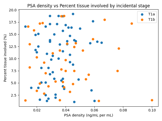 Figure 1. Distribution of ISUP Grade Groups by incidental stage (T1a vs T1b).
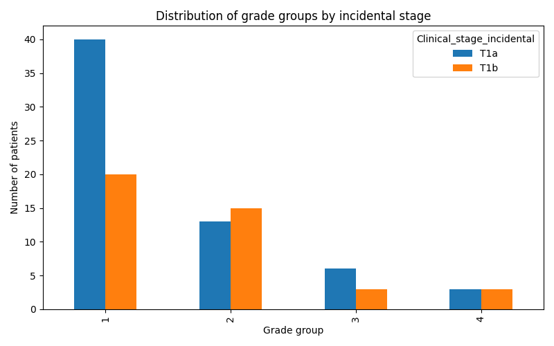 Figure 2. Exploratory relationship between PSA density and recorded percentage of TURP chips involved by cancer, colored by incidental stage. Note: The scatter plot is retained in its original form for continuity of the manuscript visuals. Stage assignment and all statistical analyses were based on the pathology-recorded thresholds of <5% for T1a and ≥5% for T1b; percentage tissue involvement was not modeled as an independent predictor of T1b.
For Grade Group ≥2 cancer, the exploratory model also had modest explanatory power (pseudo R²=0.064) and discrimination (AUC=0.65). Preoperative IPSS was the only variable significantly associated with clinically significant disease: each one-point increase in IPSS corresponded to an 8% increase in the odds of Grade Group ≥2 cancer (OR 1.08; p=0.024). A difference of several points in symptom score could therefore correspond to a meaningful difference in adjusted odds, but the association should not be interpreted as a deterministic relationship. Age, total PSA, prostate volume, PSAD, and percentage tissue involvement were not significantly associated with grade. The PSAD estimate for this model was rescaled to a 0.01 ng/mL/mL increment and was not statistically significant (OR 1.29; p=0.53), indicating that the PSAD association appeared more relevant to the extent of TURP tissue involved than to Grade Group in this cohort. Absolute PSA also failed to discriminate grade, which is unsurprising in a population selected to have values below 4 ng/mL and reinforces the limited sensitivity of a single low PSA value for clinically significant disease. Prostate volume and percentage tissue involvement produced odds ratios close to unity, while age did not demonstrate an independent effect. These results suggest that symptom burden may contain additional clinical information in this selected low-PSA population, but the AUC of 0.65 is insufficient for individual prediction. The association requires external validation and should be integrated with pathology, DRE, PSAD, imaging, and residual-gland assessment rather than used alone to alter biopsy or treatment decisions.
|
Table 4. Multivariable logistic regression models for predictors of stage T1b and of clinically significant (Grade ≥2) cancer. |
||||
|
Variable |
OR (Stage T1b) |
p (Stage T1b) |
OR (Clinically significant) |
p (Clinically significant) |
|
Age (per year) |
1.01 |
0.83 |
1.01 |
0.67 |
|
PSA (ng/mL) |
0.17 |
0.041 |
0.78 |
0.73 |
|
Prostate volume (per mL) |
1.04 |
0.17 |
1.00 |
0.86 |
|
PSA density (per ng/mL/mL) |
~3.07 × 10^42 |
0.050 |
~7.97 × 10^10 |
0.53 |
|
Percent chips involved |
0.97 |
0.53 |
0.98 |
0.72 |
|
Pre-operative IPSS |
1.04 |
0.18 |
1.08 |
0.024 |
PSAD was the strongest PSA-related factor associated with T1b disease, although the association was borderline and the adjusted model was statistically unstable. The biological rationale is plausible: PSAD expresses PSA relative to gland volume, so a PSA value that appears reassuring in isolation may be disproportionate when the gland is small. A man with PSA 3.5 ng/mL and a 20-mL gland has a PSAD of 0.175 ng/mL/mL, whereas a man with the same PSA and a 100-mL gland has a PSAD of 0.035 ng/mL/mL. Both men have PSA below the conventional 4-ng/mL threshold, yet their PSA output relative to gland size is very different. In biopsy and MRI cohorts, PSAD generally discriminates clinically significant disease better than PSA alone, and a threshold around 0.15 ng/mL/mL is commonly used to support further evaluation [5, 6, 7, 8]. However, the optimal threshold varies with gland size, population, MRI findings, and the diagnostic pathway, and some contemporary studies support lower or volume-adjusted thresholds. PSAD values in our cohort were low overall, with a mean of 0.037 ng/mL/mL; therefore, the observed continuous association occurred within a range well below 0.15 and must not be interpreted as validation of that cutoff. It instead suggests that relative differences in PSA production may remain informative even within a low range. The inverse adjusted association between absolute PSA and T1b disease is counterintuitive and does not imply that lower PSA causes or reliably predicts greater tumor extent. It may reflect the deliberately narrow PSA range, mathematical coupling among PSA, prostate volume, and PSAD, and the modest number of events. It may also reflect benign PSA production in larger glands or relatively low PSA secretion by some tumors, but these mechanisms were not directly measured. PSA is not cancer-specific: benign enlargement, inflammation, urinary retention, catheterization, and recent instrumentation can raise PSA, while some tumors may produce relatively little PSA. Absolute PSA should therefore be interpreted together with gland volume, PSAD, DRE findings, symptoms, recent clinical events, and imaging where available. In resource-limited settings, calculation of PSAD from a routinely obtained PSA and ultrasound-estimated volume is feasible, but the result should guide closer assessment rather than be used as a stand-alone diagnostic test. Because total PSA values in this cohort were low and overlapped with the range commonly encountered in benign prostatic hyperplasia, a single total PSA measurement lacked sufficient sensitivity for clinically significant incidental cancer and should not be interpreted in isolation.
Preoperative LUTS severity, measured by IPSS, was independently associated with Grade Group ≥2 disease. Each one-point increase in IPSS was associated with an 8% increase in the odds of clinically significant cancer in the adjusted model. This is a novel and hypothesis-generating observation because most studies of incidental prostate cancer emphasize age, PSA, DRE findings, prostate size, and specimen characteristics rather than symptom scores. Several explanations are possible. More severe symptoms may reflect greater transition-zone distortion, coexisting benign obstruction, inflammation, or tumor-related effects on the bladder outlet. Tumors situated within or adjacent to the transition zone could contribute directly to obstruction or irritative symptoms, while chronic outlet obstruction may also lead to earlier surgical intervention and more extensive tissue sampling. A high IPSS may identify men with longer-standing symptoms, repeated retention, or delayed evaluation, all of which may coexist with cancer without being caused by it. Nevertheless, IPSS is not a cancer-specific instrument, and symptoms caused by benign prostatic hyperplasia are common in men without malignancy. The association could also be influenced by unmeasured factors such as bladder dysfunction, prostate configuration, median lobe enlargement, medication use, or duration of symptoms. The modest model discrimination (AUC 0.65) confirms that IPSS alone is inadequate for individual risk prediction. The finding should therefore not be used as a stand-alone indication for biopsy or definitive treatment. Rather, severe symptoms in a man with low PSA should encourage careful integration of DRE, prostate volume, PSAD, imaging, and pathological assessment. Prospective studies should record symptom subdomains, uroflowmetry, post-void residual volume, retention history, and anatomical features to determine whether the association is reproducible and biologically meaningful.
Traditional factors such as age, absolute PSA, and prostate volume did not reliably distinguish more extensive or higher-grade disease once the selected low-PSA, cancer-positive population was considered. Older age is associated with the overall probability of prostate cancer and with iPCa detection in some TURP series [2, 11, 15], but age may have limited ability to discriminate stage or grade after cancer has already been identified. The restricted age distribution of men selected for surgery may also reduce the apparent association. Prostate size and the quantity of tissue removed have an ambiguous relationship with incidental staging. Larger glands may yield more tissue for pathological examination, increasing the chance of finding a small focus, but a large resection may dilute the percentage of tissue occupied by that focus and favor a T1a classification. Conversely, a limited resection can classify a small absolute focus as T1b when it represents ≥5% of the specimen [11, 12]. The ratio of resected tissue weight to estimated total gland weight may therefore be more informative than specimen weight alone because it describes the fraction of the gland sampled. This ratio could help distinguish a specimen in which a small proportion of a very large gland was removed from one in which most of a smaller transition zone was resected. It still would not measure unsampled peripheral-zone cancer, but it would improve interpretation of the pathological burden within the context of the operation. We could not analyze this ratio because resected weights were not consistently recorded. The absence of these data limits interpretation of tissue burden and supports prospective recording of preoperative gland volume, resected weight, sampling fraction, number of chips or blocks, and standardized pathological processing in future studies. Central pathology review would also allow consistent assessment of Grade Group, cribriform morphology, perineural invasion, lymphovascular invasion, and other adverse features that were not uniformly available in the retrospective reports. Across published incidental-cancer studies, higher PSAD, a lower free-to-total PSA ratio, advanced age, smaller prostate volume, and suspicious multiparametric MRI findings have been proposed as markers of clinically significant disease; however, free PSA and MRI were unavailable in the present cohort, and age and prostate volume were not independent predictors here. These factors should therefore be regarded as complementary elements of residual-gland assessment rather than universally validated thresholds.
No patient in this cohort had usable preoperative multiparametric MRI data, and PI-RADS scores or radiological features could not be correlated with stage or grade. MRI was not routinely available or affordable during the study period, which reflects the realities of the setting rather than an analytical omission that can be corrected retrospectively. Before TURP, a relatively high PSA in a small gland or a PSAD that appears disproportionate should prompt closer evaluation for occult cancer. Where resources permit, this may include repeating PSA after treating reversible causes, careful DRE, multiparametric MRI, and targeted plus systematic biopsy. After an incidental diagnosis, MRI may help assess the residual peripheral zone, identify suspicious lesions, guide confirmatory biopsy, and refine treatment planning, although MRI-negative clinically significant disease remains possible [20, 21]. The absence of imaging data also prevents us from determining whether any of the higher-grade cancers had pre-existing radiological features that might have altered the initial surgical pathway. Future prospective studies should incorporate standardized ultrasound measurements, MRI where available, and explicit documentation of reasons for proceeding directly to TURP.
The management implications are substantial because 43 patients had Grade Group ≥2 disease, while 60 had Grade Group 1 cancer. Absolute PSA did not discriminate grade reliably in this cohort, so post-TURP management should not be based solely on the preoperative PSA or on symptomatic improvement after resection. TURP removes mainly transition-zone tissue and is not an oncological procedure; residual cancer may remain in the transition zone or peripheral gland even when the postoperative urinary symptoms improve. Follow-up should therefore be structured. A post-TURP PSA baseline may be established after postoperative inflammation has resolved and repeated at approximately 3, 6, and 12 months, followed by annual testing when stable. PSA kinetics should be interpreted in relation to the reduced gland volume, and a rising PSA, concerning PSAD in the residual gland, abnormal DRE, Grade Group ≥2 disease, or other adverse features should prompt MRI, confirmatory systematic and targeted biopsy, and appropriate staging where available. The intensity of surveillance should reflect grade, stage, treatment intent, life expectancy, and access to care. Management must be individualized according to age, comorbidities, functional status, life expectancy, cancer grade, residual disease assessment, metastatic status, and patient preferences. Older or frail men with limited Grade Group 1 disease may be suitable for active surveillance or watchful waiting, particularly when confirmatory assessment does not identify higher-grade disease. Active surveillance requires a defined monitoring plan and differs from passive loss to follow-up. Fit patients with localized Grade Group ≥2 disease should be evaluated for potentially curative radical prostatectomy or radiotherapy, while selected men may be considered for surveillance only after careful assessment of volume and risk. Suspected metastatic disease requires appropriate imaging and systemic treatment planning [17, 18, 19, 22]. Histological upgrading in a subsequent biopsy or radical prostatectomy specimen can alter treatment, but no consistent radical prostatectomy pathology dataset was available in this study. In the present retrospective records, serial PSA values, treatment selection, recurrence, metastasis, survival, confirmatory biopsies, and radical prostatectomy pathology were not documented consistently enough for a valid outcome or concordance analysis. We therefore do not report anecdotal progress as study findings and have not invented upgrading, downstaging, or definitive-treatment outcomes. Establishing a prospective iPCa registry would permit evaluation of treatment patterns, adherence to PSA surveillance, pathological concordance, biochemical recurrence, metastasis-free survival, and cancer-specific survival in this setting.
Several limitations constrain interpretation. First, the retrospective case-only design is susceptible to selection and information bias and cannot estimate the incidence of iPCa among all men undergoing TURP because the denominator of eligible procedures was not available. The findings therefore describe the distribution and predictors within cancer-positive cases rather than the risk of discovering cancer in a general TURP population. Selection may also have been influenced by referral patterns, surgical eligibility, completeness of PSA testing, and the availability of pathology reports. Second, the sample size was modest relative to the number of variables entered into the regression models, the AUC values were low, and uncertainty around the PSAD estimates was substantial. PSA, prostate volume, and PSAD are mathematically related, which may have contributed to coefficient instability, suppression effects, and the inverse PSA finding. Internal resampling or external validation was not available, and the models should not be treated as clinical calculators. Third, pathological stage was based on the percentage of resected TURP tissue involved and not on whole-gland tumor volume. Resected tissue weight and the resection-to-gland ratio were not consistently recorded, and there was no central pathology review or uniform reporting of other adverse histological features. Fourth, preoperative MRI, postoperative imaging, systematic confirmatory biopsy, and high-resolution archival photomicrographs were unavailable, so radiological-pathological correlation, review of representative microscopic morphology, and assessment of residual-gland disease were impossible. The absence of images reflects the lack of a central digital pathology archive and should not be addressed by inserting non-patient or fabricated photomicrographs. Fifth, treatment, serial PSA, radical prostatectomy pathology, upgrading or downstaging, recurrence, metastasis, and survival were not systematically captured. Incomplete follow-up may be particularly important in a resource-limited setting where travel, cost, and access influence surveillance. Finally, the restriction to PSA <4 ng/mL improves focus but limits generalizability to men with higher PSA values and may produce relationships that differ from those in unselected TURP cohorts. These limitations make the results exploratory and highlight the need for prospective studies with complete TURP denominators, standardized specimen processing, tissue-weight measurements, imaging, confirmatory assessment of the residual gland, and longitudinal follow-up. Such studies should prespecify parsimonious models, avoid redundant mathematically coupled predictors, and validate any PSAD or symptom-score thresholds before clinical implementation.
Ethical approval was obtained from the Cameroon Baptist Convention Health Services Institutional Review Board (Approval Number: IRB2025-17, dated February 22, 2025). Administrative authorization was granted by the management of Nkwen Baptist Hospital and Regional Hospital Bamenda. The institutional review board waived individual informed consent because this was a retrospective analysis of de-identified medical records.
Human ethics and consent to participate declarations
This study involved retrospective review of human-participant records. All procedures were conducted in accordance with the ethical standards of the institutional and/or national research committee and with the 1964 Declaration of Helsinki and its later amendments. No identifiable individual patient information was included in the analysis or manuscript.
Consent to publish
Not applicable. This manuscript contains no identifiable individual person’s data, images, or videos.
Funding declaration
This research received no specific grant from any funding agency in the public, commercial, or not-for-profit sectors.
Authors’ contributions
Conceptualization: TTNE. Methodology: TTNE, FYT, MLM, AKE. Investigation and data collection: FYT, MLM, AKE, MDCB. Formal analysis: MLM, TTNE. Resources and supervision: TTNE, LOM, FFA. Writing—original draft: TTNE, MLM, MDCB. Writing—review and editing: all authors. Validation: LOM, FFA. All authors read and approved the final manuscript.
Acknowledgements
Special thanks to the administration of Nkwen Baptist Hospital and Regional Hospital Bamenda for authorizing us to use their databases and medical records for the sake of this study. Furthermore, our sincere thanks go to all the hospital staff that played a role in facilitating the study, whether through logistical support, patient coordination, or assistance in data collection. Your cooperation and dedication were vital to the smooth execution of the research.
Use of AI-assisted technologies
During manuscript revision, an AI-assisted language tool was used to support language editing, consistency checks, and preparation of revised graphical material. The authors critically reviewed and edited all outputs and take full responsibility for the accuracy, originality, and integrity of the final manuscript.
- Ozbey C, Oztorun K: Frequency of incidental cancer in transurethral prostate resection materials and our clinical approach to these patients: a retrospective file scan. Kafkas J Med Sci 2019, 9(1): 11-16.
- Sayar HC, Ekmekçi S, Çakmak Ö, Küçük Ü: Incidental prostate adenocarcinoma in prostate transurethral resections: our eight-year experience. J Urol Surg 2020, 7(2): 88-91.
- Janjua TK, Yousuf MA, Iqbal MT, Memon SM, Abdullah A, Faridi N, Irfan M: Incidental finding of prostate cancer in transurethral resection of prostate specimens: a retrospective analysis from a tertiary care hospital in Pakistan. Pan Afr Med J 2021, 39: 20.
- Raphael JE, Ekeke ON: T1a/T1b transitional zone prostate cancer detection rates in patients who had transurethral resection of the prostate for clinically diagnosed benign prostatic enlargement in Southern Nigeria. Eur J Health Sci 2022, 7(2): 1-7.
- Björnebo L, Discacciati A, Abbadi A, Falagario UG, Engel JC, Vigneswaran HT, Jäderling F, Grönberg H, Eklund M, Lantz A, et al: Prostate-specific antigen density as a selection tool before magnetic resonance imaging in prostate cancer screening: an analysis from the STHLM3MRI randomized clinical trial. Eur Urol Focus 2025, Epub ahead of print.
- Robinson E, Kinsella N, Ap Dafydd D, Shur J, Sohaib A, Hazell S, Bassett P, Kumar P, Mayer E, Cahill D, et al: Prostate-specific antigen density and clinically significant prostate cancer: the influence of prostatic volume. Prostate 2025, 85(8): 784-791.
- Wen J, Liu W, Shen X, Hu W: PI-RADS v2.1 and PSA density for the prediction of clinically significant prostate cancer among patients with PSA levels of 4–10 ng/mL. Sci Rep 2024, 14(1): 6570.
- Liu Z, Yu Y, Hu R, Jian T, Yu K, Lu J: Effect of prostate volume on the predictive value of prostate-specific antigen density for prostate cancer. Transl Androl Urol 2025, 14(1): 70-80.
- Bendari A, Bendari A, Zhong X, Geetha SD, Al-Refai R, Sham S, Mohammed H, Anjali F, Unger P: Hemoglobin-Albumin-Lymphocyte-Platelet (HALP) score as a predictive value of incidental prostate cancer for patients going for transurethral resection of the prostate (TURP): a single-center study. Cureus 2024, 16(4): e57736.
- Köllermann J, Hoeh B, Ruppel D, Smith K, Reis H, Wenzel M, Preisser F, Kosiba M, Mandel P, Karakiewicz PI, et al: The significance of the extent of tissue embedding for the detection of incidental prostate carcinoma on transurethral prostate resection material: the more, the better? Virchows Arch 2022, 481: 387-396.
- Nergiz D, Yıldırım HT, Yıldırım Ş: Incidence of incidental cancer in transurethral resection of prostate specimens: a 10-year retrospective analysis. Afr J Urol 2021, 27: 120.
- Rajab R, Fisher G, Kattan MW, Foster CS, Møller H, Oliver T, Reuter V, Scardino PT, Cuzick J, Berney DM; Transatlantic Prostate Group: An improved prognostic model for stage T1a and T1b prostate cancer by assessments of cancer extent. Mod Pathol 2011, 24(1): 58-63.
- Osegbe DN: Prostate cancer in Nigerians: facts and non-facts. J Urol 1997, 157(4): 1340-1343.
- Mohamed A, Abdullahi I, Warsame F, Mohamud H: Incidence and associated factors for incidental prostate cancer among patients who underwent surgery for benign prostatic hyperplasia: first report from Somalia. J Cancer Res Clin Oncol 2023, 149: 4041-4046.
- Varghese J, Kuruvilla PM, Mehta N, Rathore RS, Babu M, Bansal D, Pillai B, Sam MP, Krishnamorthy H: Incidentally detected adenocarcinoma prostate in transurethral resection of prostate specimens: a hospital-based study from India. Asian Pac J Cancer Prev 2016, 17(4): 2255-2258.
- Perera M, Lawrentschuk N, Perera N, Bolton D, Clouston D: Incidental prostate cancer in transurethral resection of prostate specimens in men aged up to 65 years. Prostate Int 2016, 4(1): 11-14.
- Bologna E, Licari LC, Franco A, Ditonno F, Manfredi C, De Nunzio C, Antonelli A, De Sio M, Leonardo C, Simone G, et al: Incidental prostate cancer in patients treated for benign prostatic hyperplasia: analysis from a contemporary national dataset. Diagnostics (Basel) 2024, 14(7): 677.
- Otto B, Barbieri C, Lee R, Te AE, Kaplan SA, Robinson B, Chughtai B: Incidental prostate cancer in transurethral resection of the prostate specimens in the modern era. Adv Urol 2014, 2014: 627290.
- Leni R, Vickers AJ, Brasso K, Montorsi F, Briganti A, Nielsen TK, Røder A, Stroomberg HV: Management and oncologic outcomes of incidental prostate cancer after transurethral resection of the prostate in Denmark. J Urol 2024, 212(5): 692-700.
- Kamrava M, Kishan AU, Margolis DJ, Huang J, Dorey F, Lieu P, Kupelian PA, Marks LS: Multiparametric magnetic resonance imaging for prostate cancer improves Gleason score assessment in favorable-risk prostate cancer. Pract Radiat Oncol 2015, 5(6): 411-416.
- Schoots IG, Nieboer D, Giganti F, Moore CM, Bangma CH, Roobol MJ: Is magnetic resonance imaging-targeted biopsy a useful addition to systematic confirmatory biopsy in men on active surveillance for low-risk prostate cancer? A systematic review and meta-analysis. BJU Int 2018, 122(6): 946-958.
- Porto JG, Blachman-Braun R, Ajami T, Zarli M, Chen R, Furtado T, Marcovich R, Parekh DJ, Shah HN: Incidental prostate cancer after holmium laser enucleation of the prostate: critical analysis of independent risk factors and impact on surgical outcomes. BJUI Compass 2024, 5(3): 374-381.
Annals of urologic oncology
p-ISSN: 2617-7765, e-ISSN: 2617-7773
 Copyright © Ann Urol Oncol. This work is licensed under a Creative Commons Attribution-NonCommercial-No Derivatives 4.0 International (CC BY-NC-ND 4.0) License.