
 Submit Manuscript
Case Report | Open Access
Prostatic Stromal Sarcoma Presenting as a Histomorphologic Outlier in Prostatic Neoplasia A Case Report
Arushi, Sufian Zaheer, Abhishek Lachyan
1Department of Pathology, Vardhman Mahavir Medical College (VMMC) & Safdarjung Hospital, New Delhi, India.
Correspondence: Zaheer Sufian (Department of Pathology, Vardhman Mahavir Medical College, VMMC & Safdarjung Hospital, New Delhi, India; Email: sufianzaheer@gmail.com).
Annals of Urologic Oncology 2026, 9: 8. https://doi.org/10.32948/auo.2026.10.21
Received: 19 Sep 2026 | Accepted: 21 Oct 2026 | Published online: 11 Nov 2026
Case presentation A 70-year-old man presented with progressive lower urinary tract symptoms and recurrent urinary retention, having undergone transurethral resection of the prostate (TURP) 10 months earlier, reported as benign prostatic hyperplasia. Ultrasonography revealed an enlarged heterogeneous prostate with an 8 × 7 mm intraprostatic lesion. Serum PSA was 5.09 ng/mL. Uroflowmetry demonstrated reduced maximum flow (9.6 mL/s) with a voided volume of 128 mL. Contrast-enhanced computed tomography revealed a large heterogeneous prostatic mass with rectal displacement and loss of fat planes with the bladder base. ¹⁸F-FDG PET/CT demonstrated necrosis and extension into the rectoprostatic space, with an indeterminate right inguinal lymph node.
Result TURP revealed a highly cellular malignant spindle-cell neoplasm exhibiting intersecting fascicles, storiform architecture, marked nuclear atypia, bizarre multinucleated cells, atypical mitoses (8–10/10 high-power fields), and coagulative necrosis. Immunohistochemistry demonstrated diffuse vimentin and focal CD34 positivity, with negativity for pan-cytokeratin, desmin, smooth muscle actin, STAT6, CD117, S100, progesterone receptor, and epithelial membrane antigen. The Ki-67 proliferation index was approximately 15%. These findings supported PSS. Molecular testing was unavailable. The patient subsequently sought treatment elsewhere and was lost to follow-up.
Conclusion PSS may initially mimic benign prostatic disease. Progressive symptoms and suspicious imaging despite previous benign histology warrant pathological reassessment. Diagnosis requires careful morphological evaluation, targeted immunohistochemistry, and exclusion of mimics. Multidisciplinary management and accurate staging are essential.
Key words Prostatic stromal sarcoma, Prostatic stromal tumor of uncertain malignant potential, Spindle-cell neoplasm, Immunohistochemistry, Prostate, Transurethral resection, Case report
The clinical presentation of PSS is often nonspecific and may include lower urinary tract symptoms, urinary obstruction, urinary retention, dysuria, hematuria, or pelvic discomfort [1, 4]. Because PSS arises from mesenchymal rather than epithelial tissue, serum prostate-specific antigen (PSA) may not be markedly elevated and cannot reliably exclude the diagnosis [1, 4]. Consequently, the initial clinical impression may be benign prostatic hyperplasia (BPH), particularly in older men presenting with progressive urinary symptoms.
Radiological findings are variable and may include a heterogeneous, lobulated, or multinodular prostatic mass with areas of necrosis and extension into adjacent pelvic structures [4, 5]. However, these features are not specific to PSS and may overlap with BPH, STUMP, leiomyosarcoma, sarcomatoid carcinoma, solitary fibrous tumor, gastrointestinal stromal tumor, and other spindle-cell neoplasms. Therefore, imaging findings must be interpreted alongside clinical features and histopathological examination.
Histopathological evaluation is central to the diagnosis of PSS. Typical malignant features include increased stromal cellularity, marked nuclear atypia, frequent mitotic figures, atypical mitoses, tumor necrosis, and, in some cases, stromal overgrowth [1, 2]. Distinguishing PSS from STUMP is particularly important because of differences in their malignant potential and clinical implications. Immunohistochemistry assists in evaluating the differential diagnosis by identifying or excluding epithelial, smooth-muscle, neural, and other mesenchymal lineages. However, no single immunohistochemical marker is specific for PSS; findings must be interpreted in conjunction with the tumor morphology and clinical context [1, 2].
Diagnosis may be especially challenging in fragmented or limited transurethral resection of the prostate (TURP) specimens. Sampling of a heterogeneous lesion may fail to capture its most atypical or malignant component, potentially complicating interpretation. When symptoms persist or progress despite treatment for presumed BPH, particularly when subsequent imaging reveals a suspicious mass, reassessment of the original histopathological material and repeat tissue sampling should be considered where clinically indicated.
The rarity of PSS limits the availability of high-quality evidence regarding its molecular characteristics, treatment, and prognosis. Published evidence consists predominantly of case reports and small clinicopathological series, and management is generally individualized according to tumor extent, resectability, and the patient's clinical condition [1, 4, 6, 7]. Furthermore, molecular investigations have demonstrated heterogeneity among tumors historically classified as specialized prostatic stromal neoplasms, highlighting the limitations of relying exclusively on historical morphological classifications [8].
We present the case of a 70-year-old man with progressively worsening lower urinary tract symptoms and a large, locally extensive prostatic mass identified approximately 10 months after an initial TURP specimen had been interpreted as BPH. Repeat TURP and subsequent histopathological and immunohistochemical evaluation supported a diagnosis of PSS. This case highlights the diagnostic challenges associated with an initially benign pathological interpretation followed by clinical and radiological progression. It also emphasizes the importance of clinicopathological correlation, careful evaluation of spindle-cell neoplasms, and transparent documentation of diagnostic limitations and unavailable follow-up information.
The patient had undergone transurethral resection of the prostate (TURP) approximately 10 months before the current presentation. Histopathological examination of the original TURP specimen had been reported as benign prostatic hyperplasia (BPH). In view of subsequent clinical progression, the available original slides and paraffin-embedded tissue blocks were retrospectively reviewed. The reviewed material showed predominantly benign prostatic hyperplasia without definite evidence of the malignant spindle-cell proliferation identified in the subsequent specimen. Given the fragmented nature of the original TURP material, sampling limitation could not be excluded as a possible explanation for the discrepancy.
On current examination, digital rectal examination revealed grade III prostatomegaly. The prostate was non-tender and non-nodular, with free overlying mucosa.
Transrectal ultrasonography (TRUS) demonstrated an enlarged prostate with heterogeneous echotexture and a well-circumscribed intraprostatic lesion measuring approximately 8 × 7 mm. The total prostate volume was not documented in the available clinical records. Serum prostate-specific antigen (PSA) was 5.09 ng/mL, within the age-specific reference range used by the treating institution for a 70-year-old man (Figure 1).
Uroflowmetry demonstrated a voided volume of 128 mL and a maximum urinary flow rate (Qmax) of 9.6 mL/s, with an interrupted and flattened flow curve. Because the voided volume was relatively low, the Qmax value was interpreted cautiously, as a low voided volume may limit the reliability of uroflowmetric assessment. The postvoid residual urine volume was not documented in the available records (Figure 2).
In view of persistent and progressive symptoms despite the previous benign pathological diagnosis, further radiological evaluation was undertaken. Contrast-enhanced computed tomography (CECT) of the pelvis demonstrated a large, heterogeneously enhancing mass involving the prostate, with posterior and right lateral extension. The lesion produced mass effect on the rectum, resulting in left posterolateral displacement and compression. There was loss of the intervening fat planes between the mass and the urinary bladder base, raising concern for possible local invasion. Possible involvement of the levator ani muscles was also reported (Figure 3).
An enlarged right inguinal lymph node measuring approximately 12 × 8 mm was identified on imaging. The node was considered indeterminate. Subsequent whole-body ^18F-fluorodeoxyglucose positron emission tomography/computed tomography (^18F-FDG PET/CT) demonstrated a large, lobulated, heterogeneously enhancing soft-tissue mass arising predominantly from the posterior aspect of the right prostatic lobe. The lesion contained areas of necrosis and extended into the rectoprostatic space, raising concern for an aggressive prostatic neoplasm. The available staging examination did not demonstrate definitive evidence of metastatic involvement of the right inguinal lymph node or definite distant metastatic disease (Figure 4). The node was not biopsied. The available records did not establish whether subsequent dedicated radiological follow-up of the lymph node was performed; therefore, its later status could not be determined.
In view of the progressive urinary symptoms and suspicious radiological findings, repeat TURP was performed, and the resected tissue was submitted for histopathological examination.
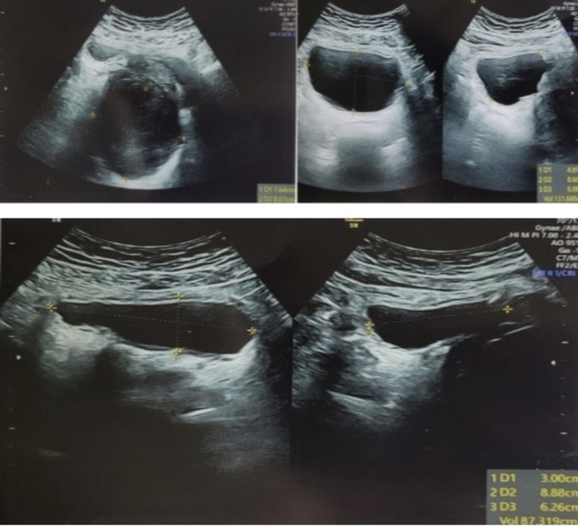 Figure 1. Ultrasonography of the prostate showing enlargement with heterogeneous echotexture and a well-defined 8 × 7 mm intraprostatic lesion.
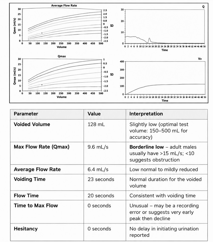 Figure 2. Uroflowmetry showing low voided volume (128 mL), Qmax 9.6 mL/s, and a flattened, interrupted flow curve, suggestive of bladder outlet obstruction.
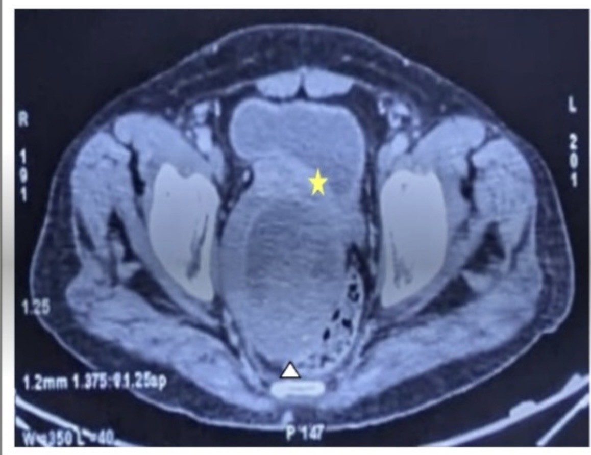 Figure 3. CE-CT showing a large heterogeneously enhancing prostatic mass infiltrating the urinary bladder (asterisk), with rectal displacement and loss of intervening fat planes (arrowhead).
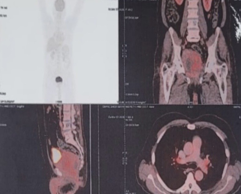 Figure 4. ¹⁸F-FDG PET/CT showing a large, lobulated prostatic mass with heterogeneous uptake and areas of necrosis, extending into the rectoprostatic space. The right inguinal lymph node was indeterminate, without definitive evidence of nodal or distant metastasis on the available staging examination.
Microscopic examination revealed a highly cellular spindle-cell neoplasm arranged predominantly in intersecting fascicles and areas of storiform architecture. The tumor cells exhibited moderate-to-marked nuclear pleomorphism, hyperchromasia, and occasional bizarre multinucleated forms. Mitotic activity was increased, with approximately 8–10 mitotic figures per 10 high-power fields, including atypical mitotic figures. Foci of coagulative tumor necrosis were also identified (Figure 5).
The malignant spindle-cell proliferation was morphologically distinct from the benign prostatic glandular and stromal components present in the specimen. Areas of pronounced stromal cellularity and atypia were conspicuous. The combination of marked cytological atypia, increased mitotic activity, atypical mitoses, and tumor necrosis supported the interpretation of a malignant mesenchymal neoplasm.
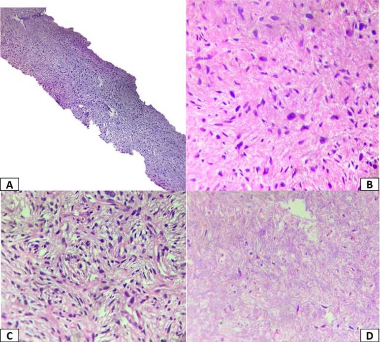 Figure 5. Histopathological features. (A) Low-power H&E (100×); (B) High-power H&E showing atypia and focal myxoid change (400×); (C) High-power H&E showing storiform pattern (400×); (D) Necrosis (400×).
The immunohistochemical findings were interpreted in conjunction with the histomorphological features rather than on the basis of any individual marker. Diffuse vimentin expression supported mesenchymal differentiation but was nonspecific. Focal CD34 positivity was interpreted in the context of the overall morphological and immunohistochemical profile. Negative panCK and EMA staining argued against an epithelial malignancy, including sarcomatoid carcinoma. The absence of desmin and SMA expression did not support smooth-muscle differentiation. S100 negativity made neural or melanocytic differentiation less likely. Negative CD117 reduced the likelihood of a gastrointestinal stromal tumor, while negative STAT6 staining argued against solitary fibrous tumor. The absence of PR expression was considered non-exclusionary and was interpreted alongside the other findings (Figure 2).
Detailed technical information regarding the antibodies, including manufacturer, clone, catalogue number, and dilution, was not available in the records accessible to the authors. This limitation is acknowledged in the interpretation of the immunohistochemical findings.
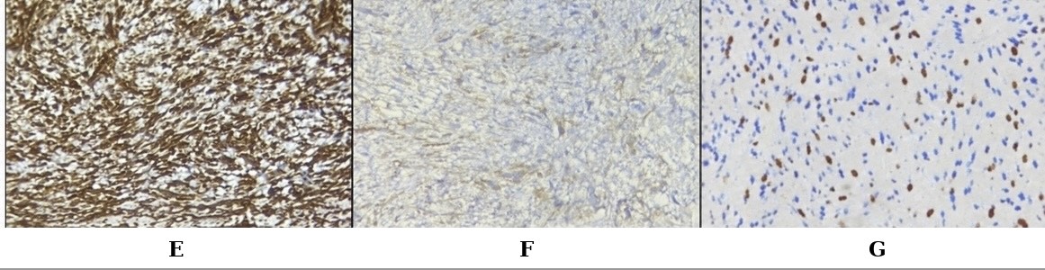 Figure 6. Immunohistochemical findings. (E) Vimentin showing diffuse cytoplasmic positivity (400×); (F) CD34 showing focal positivity (400×); (G) Ki-67 proliferation index: 15% (400×).
|
Table 1. The immunohistochemical findings. |
|
|
Marker |
Result |
|
Vimentin |
Diffuse positive |
|
CD34 |
Focal positive |
|
Pan-cytokeratin (panCK) |
Negative |
|
Desmin |
Negative |
|
Smooth muscle actin (SMA) |
Negative |
|
STAT6 |
Negative |
|
CD117 (c-KIT) |
Negative |
|
S100 |
Negative |
|
Progesterone receptor (PR) |
Negative |
|
Epithelial membrane antigen (EMA) |
Negative |
|
Ki-67 proliferation index |
Approximately 15% |
STUMP was considered because it belongs to the spectrum of specialized prostatic stromal neoplasms. However, the marked cytological atypia, frequent mitoses, atypical mitotic figures, and coagulative necrosis favored a malignant stromal neoplasm rather than STUMP [1, 2].
Leiomyosarcoma was considered because of the intersecting fascicular spindle-cell architecture. However, the absence of desmin and SMA expression did not support smooth-muscle differentiation. Sarcomatoid carcinoma was considered because of the prostatic location and spindle-cell morphology; however, negative panCK and EMA staining argued against this diagnosis.
Solitary fibrous tumor was considered because of the spindle-cell morphology and focal CD34 expression. Nevertheless, the absence of STAT6 expression and the markedly atypical morphology did not support this diagnosis. Gastrointestinal stromal tumor was also considered because of the anatomical relationship between the prostate, rectum, and adjacent pelvic structures. Negative CD117 staining and the overall morphology made this diagnosis less likely. DOG1 testing was not performed.
The final diagnosis was prostatic stromal sarcoma, based on the combined histomorphological features and immunohistochemical findings, with the limitations of the available ancillary testing acknowledged.
Consequently, treatment response, recurrence status, subsequent metastatic progression, and overall clinical outcome could not be assessed. The subsequent status of the right inguinal lymph node also remained undetermined because follow-up imaging could not be verified from the available records.
The clinical presentation was characterized by progressively worsening lower urinary tract symptoms and intermittent urinary retention. These symptoms are nonspecific and are commonly associated with benign prostatic hyperplasia (BPH), particularly in older men. The previous TURP specimen had been interpreted as BPH, further complicating the diagnostic assessment. Retrospective review of the available original slides and tissue blocks did not demonstrate definite evidence of the malignant spindle-cell proliferation identified in the subsequent specimen. Although sampling limitation may have contributed to the discrepancy, the available findings do not establish whether the sarcoma was initially unsampled or developed subsequently. This distinction is important because a previous benign pathological diagnosis should not automatically be considered erroneous when later specimens demonstrate a different pathological process.
The serum prostate-specific antigen (PSA) level was 5.09 ng/mL, within the age-specific reference range used by the treating institution. PSA is primarily associated with prostatic epithelial activity and may not be markedly elevated in mesenchymal malignancies such as PSS [1, 4]. Therefore, a PSA level within the expected reference range does not exclude a clinically significant prostatic sarcoma. In patients with persistent or progressive urinary symptoms, PSA findings should be interpreted alongside physical examination, radiological findings, and histopathological evaluation.
The imaging findings contributed substantially to the recognition of an aggressive prostatic neoplasm. Initial transrectal ultrasonography demonstrated an 8 × 7 mm intraprostatic lesion, whereas subsequent contrast-enhanced computed tomography (CECT) and ^18F-fluorodeoxyglucose positron emission tomography/computed tomography (FDG PET/CT) demonstrated a large, heterogeneous mass with necrosis and extension into the rectoprostatic space. CECT also showed displacement and compression of the rectum and loss of intervening fat planes with the urinary bladder base, raising concern for local invasion. These findings are compatible with the reported imaging spectrum of prostatic stromal sarcoma, which may include heterogeneous masses, necrotic areas, and extension into adjacent structures [4, 5]. However, because the exact dates of the individual imaging examinations were not available, the rate of tumor growth cannot be reliably determined. Differences in imaging modality and the possibility that the initial ultrasound did not demonstrate the full extent of the lesion must also be considered.
The uroflowmetry findings showed a maximum urinary flow rate (Qmax) of 9.6 mL/s with a voided volume of 128 mL. Although the findings were compatible with impaired urinary flow, the relatively low voided volume limits the reliability of Qmax as an isolated measure of obstruction. Accordingly, uroflowmetry was considered supportive of the patient's clinical presentation rather than independently diagnostic of the underlying malignancy.
Histopathological examination remains central to the diagnosis of PSS because its morphological features overlap with several other spindle-cell neoplasms. The present tumor exhibited intersecting fascicles and storiform areas, marked cytological atypia, bizarre multinucleated cells, frequent mitoses including atypical forms, and coagulative necrosis. These findings favored a malignant mesenchymal neoplasm over a benign or uncertain-potential stromal lesion [1, 2]. The differential diagnosis required consideration of STUMP, leiomyosarcoma, sarcomatoid carcinoma, solitary fibrous tumor, gastrointestinal stromal tumor, and other spindle-cell sarcomas.
Immunohistochemistry supported the morphological interpretation and assisted in evaluating the differential diagnosis. Diffuse vimentin expression was consistent with mesenchymal differentiation but was nonspecific. Focal CD34 positivity was also insufficient to establish the diagnosis independently, as CD34 expression can occur in different stromal and mesenchymal neoplasms. Negative pan-cytokeratin and epithelial membrane antigen staining argued against sarcomatoid carcinoma, while negative desmin and smooth muscle actin did not support leiomyosarcoma. Negative STAT6 staining argued against solitary fibrous tumor, and negative CD117 reduced the likelihood of gastrointestinal stromal tumor. S100 negativity made neural or melanocytic differentiation less likely. Nevertheless, individual negative markers cannot exclude every possible differential diagnosis, and the immunohistochemical panel must be interpreted in the context of morphology and the anatomical site [1, 2, 9].
The distinction between PSS and STUMP deserves particular emphasis. Both entities arise within the spectrum of specialized prostatic stromal neoplasms, but the presence and extent of malignant histological features are important in their classification [1, 2]. In this case, the combination of marked atypia, increased mitotic activity, atypical mitotic figures, and tumor necrosis favored PSS. However, molecular testing was not performed, and detailed technical information regarding the antibodies used was unavailable in the accessible laboratory records. The diagnosis is therefore supported by the available morphological and immunohistochemical findings but lacks molecular characterization and complete documentation of the immunohistochemical methodology.
Emerging molecular evidence indicates that tumors historically grouped under specialized prostatic stromal neoplasms may comprise biologically heterogeneous entities [8, 10]. This finding reinforces the importance of integrating histomorphology with appropriate ancillary investigations where available. Although molecular testing was not undertaken in the present case, the limitation has been explicitly acknowledged rather than assuming molecular confirmation.
The right inguinal lymph node measured approximately 12 × 8 mm and was considered indeterminate on imaging. The available staging examination did not establish definitive metastatic involvement. Because the node was not biopsied and subsequent dedicated radiological follow-up could not be verified from the available records, its later status remains unknown. Consequently, the possibility of subsequent nodal progression cannot be assessed, and the node should not be classified as metastatic on the basis of the available evidence alone. This uncertainty highlights the importance of documenting the results of subsequent investigations when available, particularly in patients with potentially aggressive malignancies.
Management of PSS remains challenging because the rarity of the tumor limits the availability of prospective studies and standardized treatment recommendations. Published evidence consists predominantly of case reports and small case series [1, 4, 6, 7, 11, 12]. Treatment decisions are generally individualized according to tumor extent, resectability, patient fitness, and multidisciplinary assessment. Surgical resection, radiotherapy, and systemic therapy have been described in selected cases, but the available evidence does not establish a universally accepted treatment regimen. In the present case, the patient sought oncology treatment at another cancer centre after diagnosis. The specific treatment administered and the subsequent clinical outcome were unavailable because the patient was lost to follow-up. Therefore, no conclusions can be drawn regarding treatment response, recurrence, or survival.
This case illustrates the importance of clinicopathological correlation when clinical progression is inconsistent with an earlier benign diagnosis. Persistent or worsening lower urinary tract symptoms, particularly when accompanied by a suspicious or enlarging prostatic mass, warrant further assessment even when the PSA level is not markedly elevated. Review of previous pathological material and repeat sampling may help clarify the diagnosis when clinically indicated. Equally important, case reports should distinguish documented findings from plausible explanations and explicitly acknowledge unavailable investigation dates, ancillary test details, treatment information, and follow-up outcomes.
Overall, the diagnosis of PSS in this patient was supported by the malignant spindle-cell morphology and the available immunohistochemical profile. The case emphasizes the diagnostic value of repeat tissue sampling and integrated pathological assessment in patients with discordant clinical, radiological, and pathological findings. It also demonstrates the limitations imposed by incomplete molecular characterization and loss to follow-up, which prevent a definitive assessment of subsequent disease behavior and outcome.
The authors received no specific funding for the preparation or publication of this case report.
Conflict of interest
The authors declare that they have no conflict of interest.
Ethics approval and consent to participate
Ethical approval was not required for this individual case report in accordance with the applicable institutional policies of Vardhman Mahavir Medical College and Safdarjung Hospital. The case was prepared in accordance with relevant institutional requirements and the principles of the Declaration of Helsinki.
Clinical trial number
Not applicable.
Consent for publication
Written informed consent was obtained from the patient for publication of this case report and the accompanying images.
Author contributions
Dr. Arushi: Case evaluation, data collection, histopathological evaluation, literature review, and manuscript preparation.
Dr. Sufian Zaheer and Dr. Abhishek Lachyan: Supervision, pathological interpretation, critical revision of the manuscript, and final approval of the manuscript.
Data availability
All relevant data pertaining to this case are included in the manuscript. Additional information may be requested from the corresponding author, subject to patient confidentiality and applicable institutional requirements.
Patient perspective
The patient's perspective was not obtained.
- Gaudin PB, Rosai J, Epstein JI: Sarcomas and related proliferative lesions of specialized prostatic stroma: a clinicopathologic study of 22 cases. Am J Surg Pathol 1998, 22(2): 148-162.
- Herawi M, Epstein JI: Specialized stromal tumors of the prostate: a clinicopathologic study of 50 cases. Am J Surg Pathol 2006, 30(6): 694-704.
- Moch H, Humphrey PA, Ulbright TM, Reuter VE, editors. WHO Classification of Tumours: Urinary and Male Genital Tumours. 5th ed. Lyon: International Agency for Research on Cancer; 2022.
- Yang W, Liu A, Wu J, Niu M: Prostatic stromal sarcoma: a case report and literature review. Medicine (Baltimore) 2018, 97(18): e0495.
- Tamada T, Sone T, Miyaji Y, Kozuka Y, Ito K: MRI appearance of prostatic stromal sarcoma in a young adult. Korean J Radiol 2011, 12(4): 519-523.
- Hodotsuka N, Suzuki Y, Suzuki K, Honda Y, Hatori T, Kondo Y: A case of complete resection of prostate stromal sarcoma after neoadjuvant chemotherapy. IJU Case Rep 2023, 6(4): 239-243.
- Ueda S, Okada K, Kanzawa M, Fukuda T, Furukawa J, Fujisawa M: A case of prostate stromal sarcoma involving the rectum. J Surg Case Rep 2020, 2020(6): rjaa165.
- Acosta AM, Sholl LM, Dickson BC, McKenney JK, Gordetsky JB, Pins MR, Marino-Enriquez A, Dong F, Dubuc AM, Cin PD, et al: Re-evaluating tumors of purported specialized prostatic stromal origin reveals molecular heterogeneity, including non-recurring gene fusions characteristic of uterine and soft tissue sarcoma subtypes. Mod Pathol 2021, 34(9): 1763-1779.
- Demicco EG, Park MS, Araujo DM, Fox PS, Bassett RL, Pollock RE, Lazar AJ, Wang WL: Solitary fibrous tumor: a clinicopathological study of 110 cases and proposed risk assessment model. Mod Pathol 2012, 25(9): 1298-1306.
- Cigliola A, Prakash G, Li R, Oualla K, Gupta S, Kamat AM, Chahoud J, Necchi A, Spiess PE: Current status and challenges in rare genitourinary cancer research and future directions. Curr Oncol Rep 2024, 26(9): 977-990.
- Rojas-Jiménez A, Otero-Garcia M, Mateos-Martin A: Stromal prostatic sarcoma: a rare tumor with rare clinical and imaging presentation. J Radiol Case Rep 2013, 7(7): 24-31.
- Addesso M, Caputo A, Zeppa P, D'Antonio A: Prostatic stromal sarcoma: report of a rare case in a young male and review of the literature. Int J Surg Case Rep 2023, 106: 108193.
Annals of urologic oncology
p-ISSN: 2617-7765, e-ISSN: 2617-7773
 Copyright © Ann Urol Oncol. This work is licensed under a Creative Commons Attribution-NonCommercial-No Derivatives 4.0 International (CC BY-NC-ND 4.0) License.